Radiology and Ultrasound
In veterinary medicine, X-rays and ultrasounds are powerful tools that grant exclusive glimpses into your beloved animal’s inner workings.
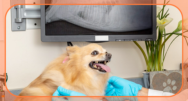
Powered by X-rays, radiology unveils intricate images by harnessing electromagnetic radiation, vividly capturing various tissue densities. These detailed snapshots reveal bones, organs, and intricate structures within your pet’s body.
Conversely, ultrasound utilizes high-frequency sound waves a probe emits, painting real-time visuals of internal organs. It interprets echoes to craft dynamic, moving images, offering invaluable insights.
Both non-invasive imaging techniques are pivotal in our diagnostic arsenal. They empower us to swiftly diagnose conditions, evaluate injuries, and vigilantly monitor your pet’s well-being without subjecting them to invasive procedures. This ensures timely and pinpoint medical interventions for your furry companion’s optimum health.
X-Rays
Your pet’s health is our top priority, and our dedication involves meticulously examining their bodily systems to ensure a comprehensive understanding of their well-being.
We unlock a detailed view of your pet’s internal functions using advanced radiological techniques like X-rays. This technology allows us to delve into crucial areas such as:
- Musculoskeletal system
- Cardiovascular system
- Respiratory system
- Gastrointestinal system
- Reproductive system
- Urinary system
We recognize the crucial role of precise diagnostics in providing optimal care for your cherished companion.
That’s why our state-of-the-art facility conducts on-site radiographs, utilizing top-notch technology to generate high-quality images swiftly.
This efficient process ensures rapid access to results, enabling our veterinary team to make well-informed decisions promptly for your pet’s well-being.
Dental X-Rays
Dental X-rays are indispensable in understanding and maintaining your pet’s oral health. While the visible portion of your pet’s teeth can tell us a lot, much of what impacts their oral and overall health lies beneath the gumline. That’s where dental X-rays come in, providing a clear and detailed look at the structures we can’t see during a routine examination.
With dental X-rays, we can:
- Examine Tooth Roots and Surrounding Bone: Dental X-rays allow us to detect hidden fractures, infections, or abscesses that might go unnoticed but could cause pain or lead to systemic issues.
- Identify Periodontal Disease: We can assess the extent of bone loss or damage caused by advanced gum disease, providing essential information for targeted treatments.
- Detect Developmental Abnormalities: These images help us identify unerupted teeth or abnormal alignment, which may require intervention to prevent further complications.
- Evaluate Tooth Decay or Resorption: Dental X-rays are crucial in pinpointing decay or resorption that cannot be seen during a visual examination, ensuring early and effective intervention.
- Monitor Post-Treatment Healing: They allow us to track healing after extractions or other dental procedures, ensuring proper recovery and long-term health.
Dental X-rays uncover these hidden issues, enabling our veterinary team to diagnose and treat oral health concerns with precision. This ensures your pet enjoys a healthier mouth and an improved quality of life. The procedure is painless and performed under anesthesia to keep your pet comfortable while providing accurate and comprehensive results.
Ultrasonography
Regarding your pet’s health, ultrasound technology becomes a crucial ally, offering detailed three-dimensional images of their internal organs through non-invasive and state-of-the-art methods.
Ultrasound gives us a more detailed view of your pet’s health than X-rays alone.
Using sound waves, ultrasound enables painless examinations of vital organs like the heart, liver, spleen, kidneys, stomach, intestines, pancreas, adrenal glands, and bladder.
When liver or kidney diseases arise, ultrasound dives deep, providing extensive insights into suspected lesions and helping us create better treatment plans.
Furthermore, ultrasonography is pivotal in guiding biopsy needles accurately to suspicious areas within organs, elevating diagnostic precision.
The meticulous ultrasound approach significantly enhances precise diagnoses, especially in cardiac cases, notably among cats.
For older cats prone to HCM-Hypertrophic Cardiomyopathy, ultrasound examinations are invaluable. This condition causes heart muscle enlargement, reduces the blood volume available to the heart, and poses severe risks.
Unlike X-rays, ultrasound stands as the primary diagnostic tool for detecting HCM. While this condition can be life-threatening without proper medical attention, many cats can be effectively managed with attentive care.
Moreover, ultrasound isn’t limited to just cardiac issues. It proves its worth in pregnancy exams, tendon evaluations, fluid collection assessments, and performing biopsies. Its versatility showcases its importance in various veterinary procedures.
Echocardiography, CT, MRI
We collaborate with Dr. Erica D. Baravick-Munsell, DVM, DACVR, and Infinity Veterinary Imaging for specialized Veterinary Radiography, Ultrasonography, and Echocardiography services.
Dr. Munsell is extensively trained in various advanced imaging techniques, including veterinary radiography, ultrasonography, echocardiography, computed tomography, magnetic resonance imaging, and nuclear medicine.
Dr. Munsell operates on a mobile basis, bringing her extensive expertise in advanced imaging techniques directly to our clinic upon request.
With her dedication to animal health and proficiency in treating small, large, and exotic animals, your pet receives top-tier specialized services conveniently at our facility.
People Also Ask:
1. Do Veterinary x-rays and ultrasounds require anesthesia?
The necessity for sedation during X-ray and ultrasound procedures depends on the specific views required.
Sedation is often unnecessary for leg X-rays as we can acquire clear images without it.
However, when focusing on pelvic X-rays, sedation becomes optimal because your dog or cat must be positioned on its back and extended legs.
This positioning is essential for obtaining precise images, as most animals tend to move, which makes it challenging to capture high-quality images without sedation.
2. How safe are veterinary X-rays and ultrasonography?
Very safe!
3. How do you determine if my pet needs X-rays, ultrasound, or both?
We use X-rays mainly for fractures, while ultrasounds are preferred for assessing internal organs like the pancreas. We aim to perform only necessary tests based on our expertise, ensuring an accurate diagnosis. We prioritize efficient and cost-effective methods to diagnose your pet’s condition promptly.
4. I already have X-rays from my current vet. Will I need to have them redone?
That depends. If the X-rays are current and have a good clear view of the problem, we can probably use them. You can bring them in or send a digital version to the clinic for us to view. The previous radiographs could also reduce the number of new views we must take.


